Ay, Ay, Eye… The anatomy of an issue.
A Wrought-Iron Project addendum
Note: This article includes graphic images essential for illustrating the AES. The specimen—hatched and deceased shortly after hatching at our facility (The Scarlet Society)—comes from our 2025 Super Wrought-Iron clutch attempt. All images and data were gathered post-mortem. Reader discretion is advised.
The Abnormal Eye Scale (AES) refers to a unique issue affecting the Wrought-Iron (WI)
Blood Python morph. This anomaly is characterized by the presence of abnormal
circumorbital scales and the spectacle, often leading to concern about the effect on the
eye and its impact on the snake. The question of whether these scales can be safely
removed was investigated. Understanding the AES issue is critical as it directly impacts
the future of the WI project, influencing breeding decisions and potentially
deterring people from it. This article aims to clarify these uncertainties by exploring the
anatomical features and implications of the AES.
To help understand this issue, I’d like to briefly describe the typical anatomy of the orbit
and circumorbital region of a blood python (Python brongersmai), so we can better analyze the key differences
between these structures in non-affected snakes vs the AES carriers.The scales surrounding the eye of a blood python are identified as
the circumorbital scales. In this species, these scales are subdivided into three sets of
scales, depending on their positions around the eye. At the top of the eye is a large
supraorbital; in the front of the eye are 2-3 preorbitals; at the back of the eye are 2-3
postoculars. One of the identifying characters of this species is the absence of suborbital
scales; instead, the bottom of the eye is bounded by one or two supralabials. (See Figure 1).The outer layer of the skin that covers the entire outer surface of a snake is called the
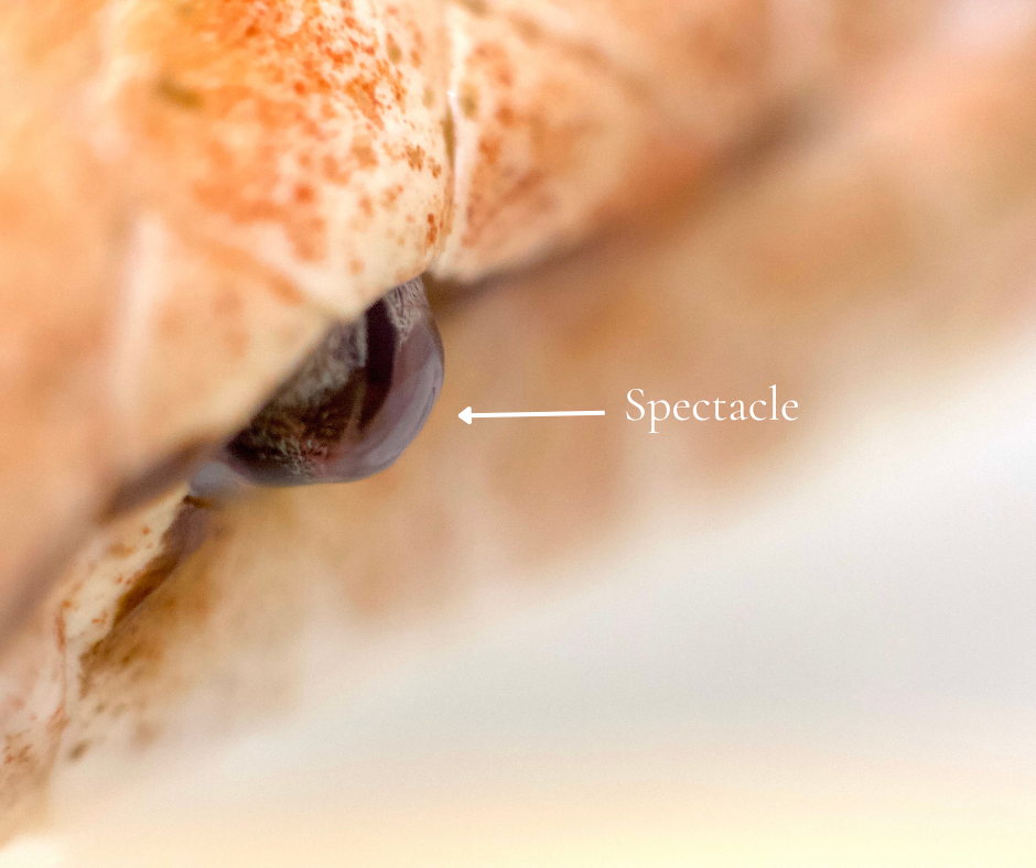
outer epidermal generation (OEG) or the stratum corneum. This layer of skin has only
four holes in it – the mouth, two nostril holes, and the cloacal vent. The OEG covers the
eye completely with a clear “contact-lens-like” structure called the spectacle. The
spectacle is part of the OEG and is completely attached around its circumference to the
OEG. (See Figure 2.)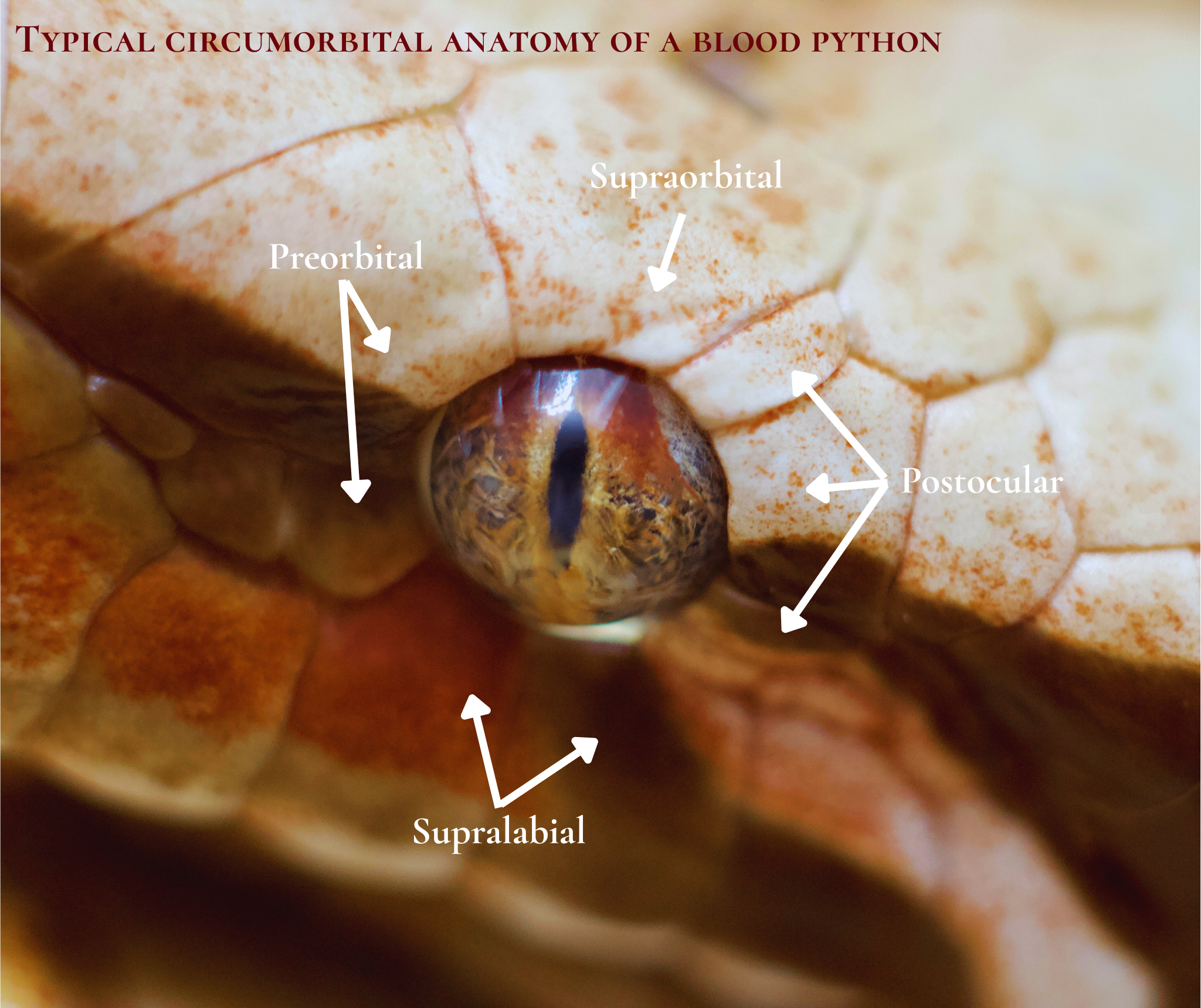
With the anatomical context established, let’s move on to the main topic. The Abnormal Eye Scale.In summary, the AES refers to the presence of aberrant scales affecting the circumorbital structures and spectacle in Wrought-Iron blood pythons, which cover and obstruct the eye to varying degrees. 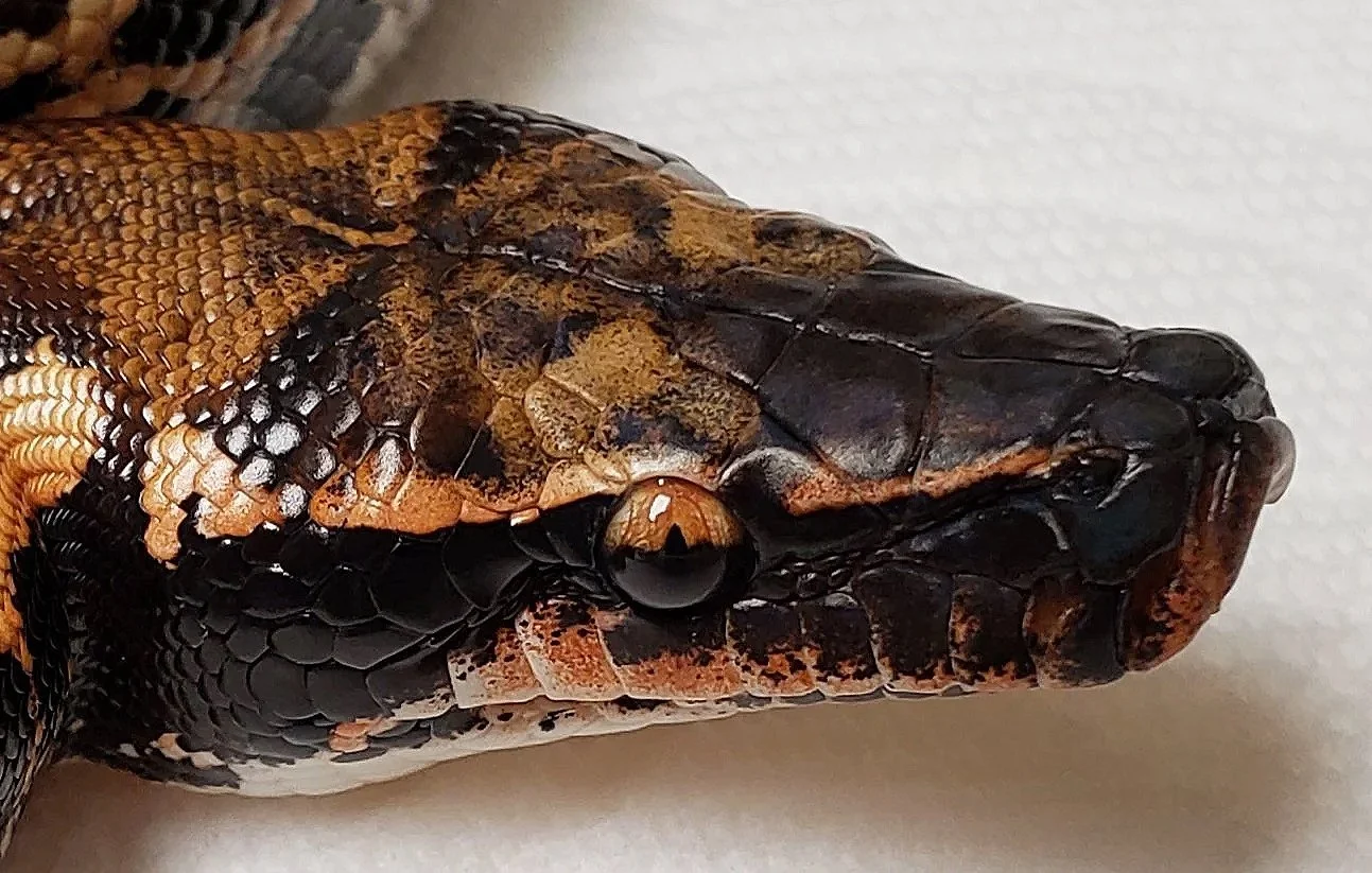
The cause of this anomaly is still unclear, and
although several hypotheses are being discussed, they remain unproven. Therefore, this article will be a descriptive approach to the observations made during the post-mortem specimen dissection.
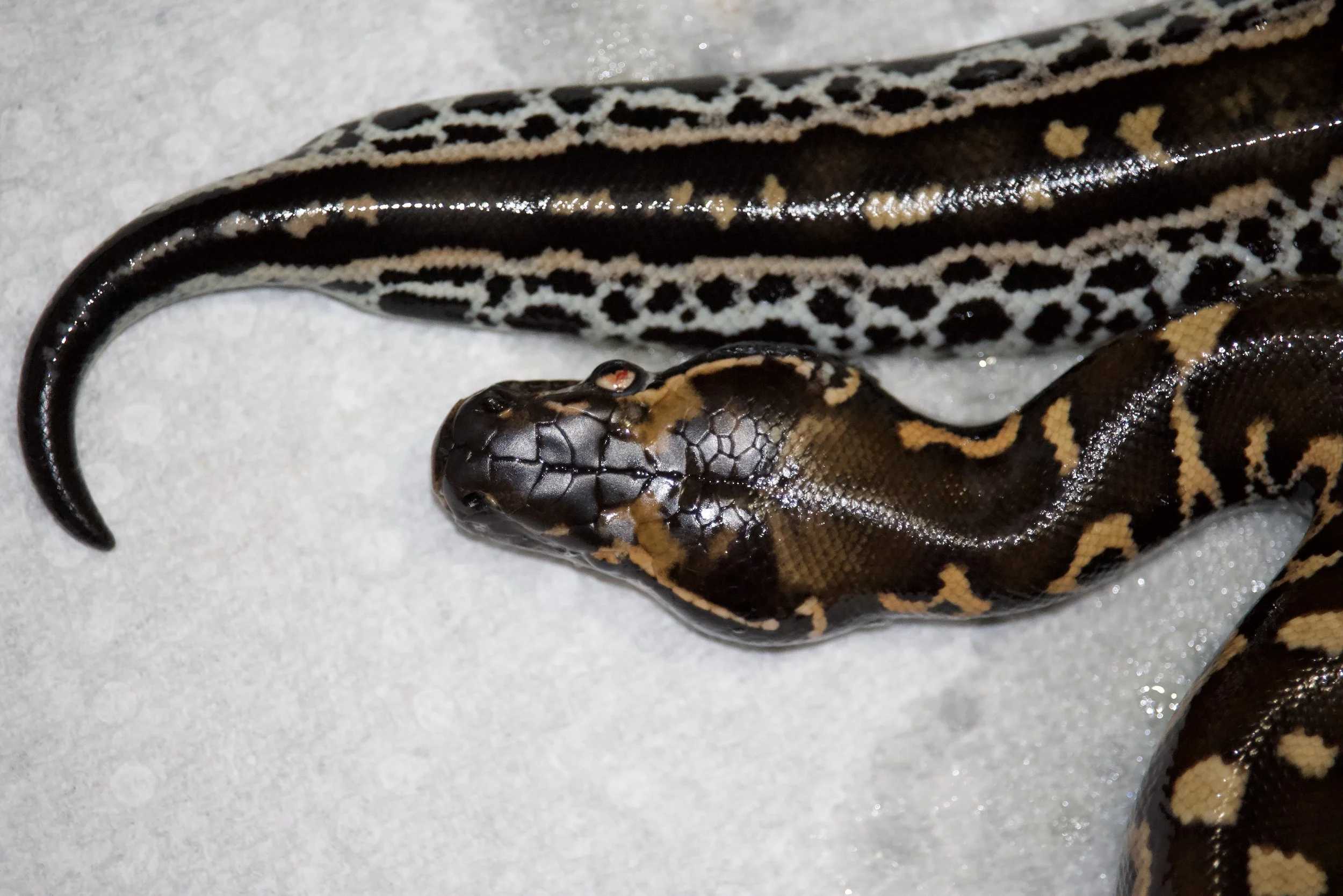
The specimen used in the dissection came from my 2025 WI to WI clutch (fig. 3). The
animal passed shortly after hatching from unknown causes, providing a unique opportunity to examine the distinct physical characteristics of the AES. Besides
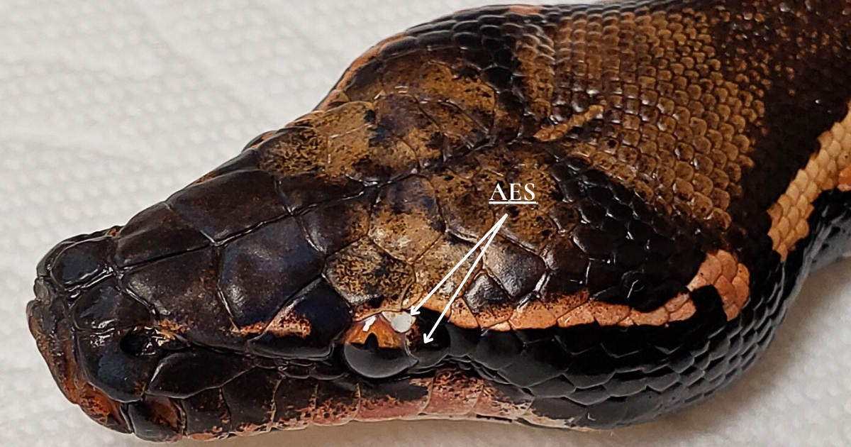
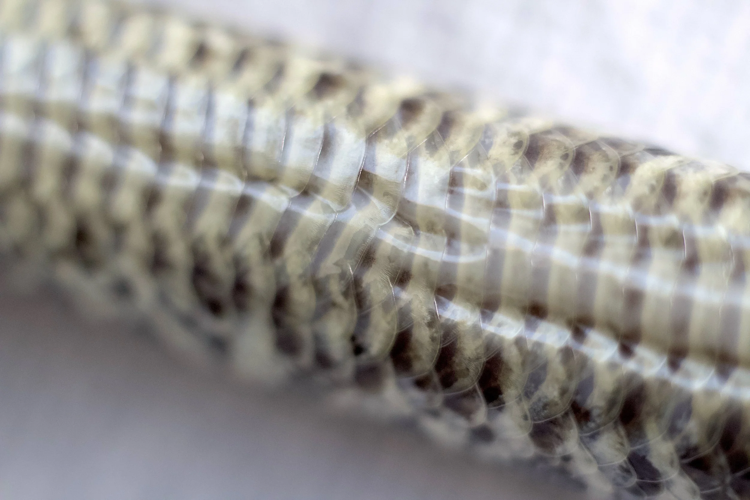
the AES (Fig. 4), all WIs from this clutch displayed other problems, such as bulging eyes (Fig. 5), jaw misshapes, and
ventral scale anomalies (Fig. 6). As of this publication, despite my efforts, all WIs from this clutch failed to thrive. They never accepted food and passed away approximately 2.5 months after hatching.
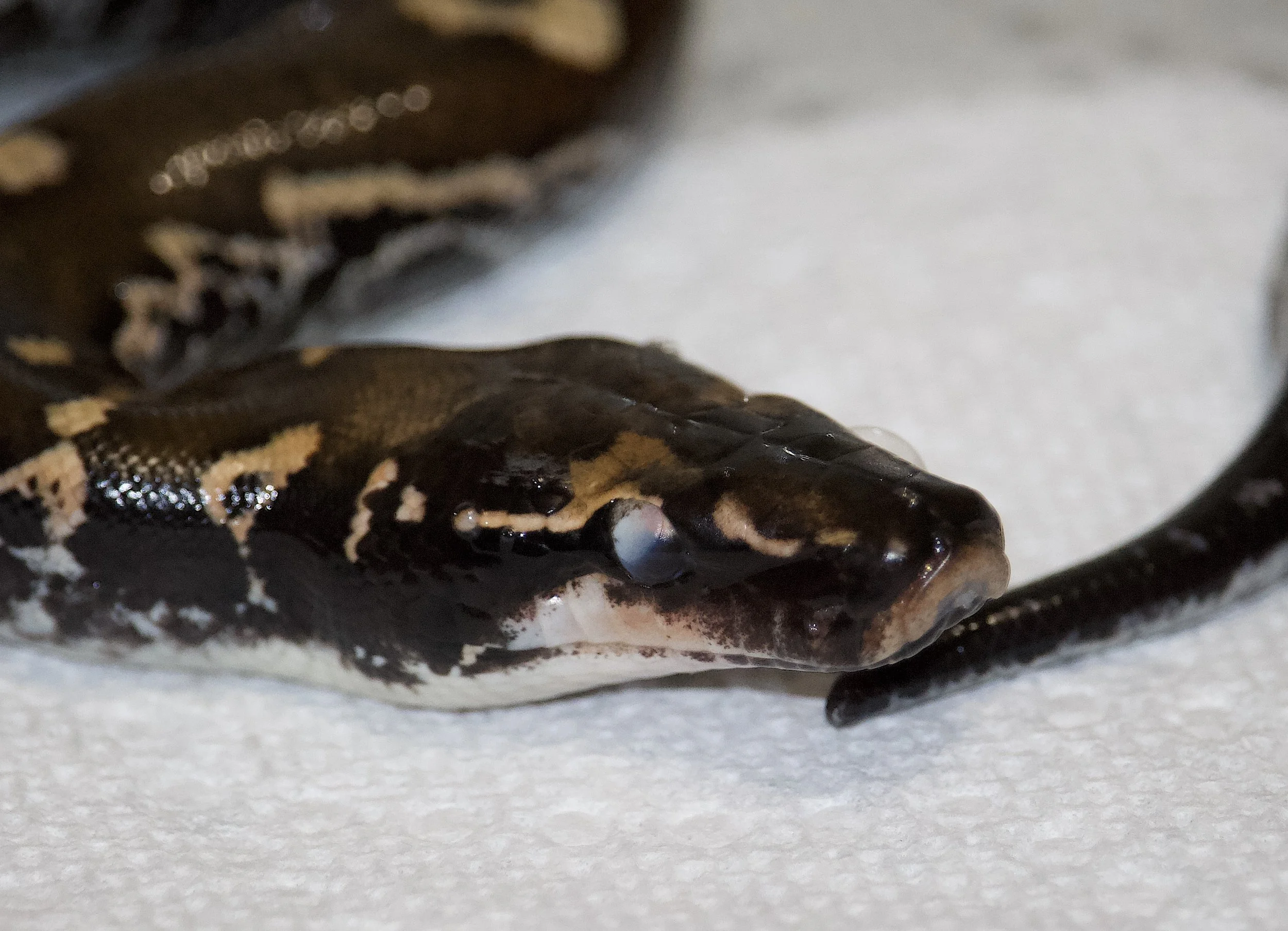
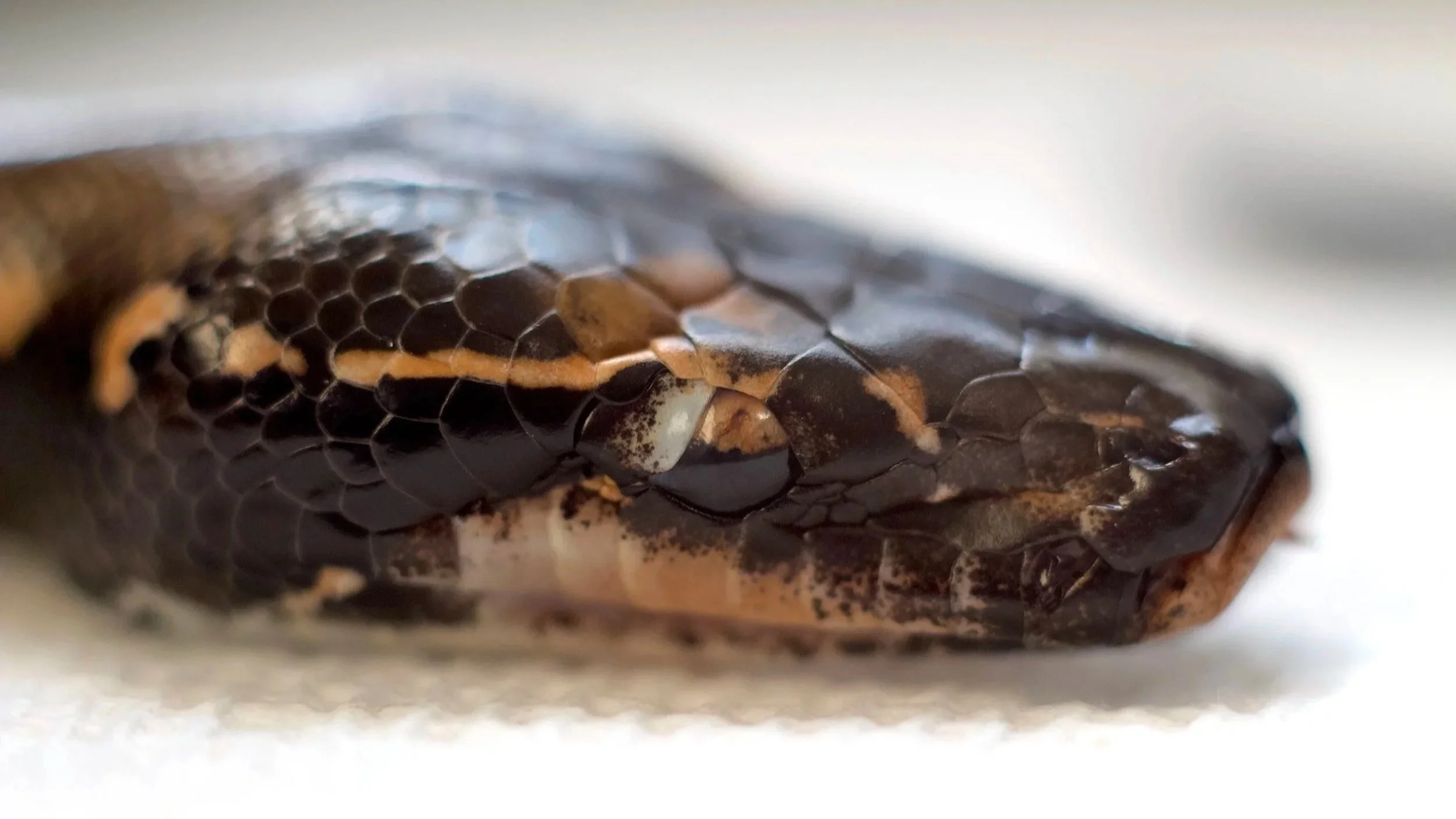
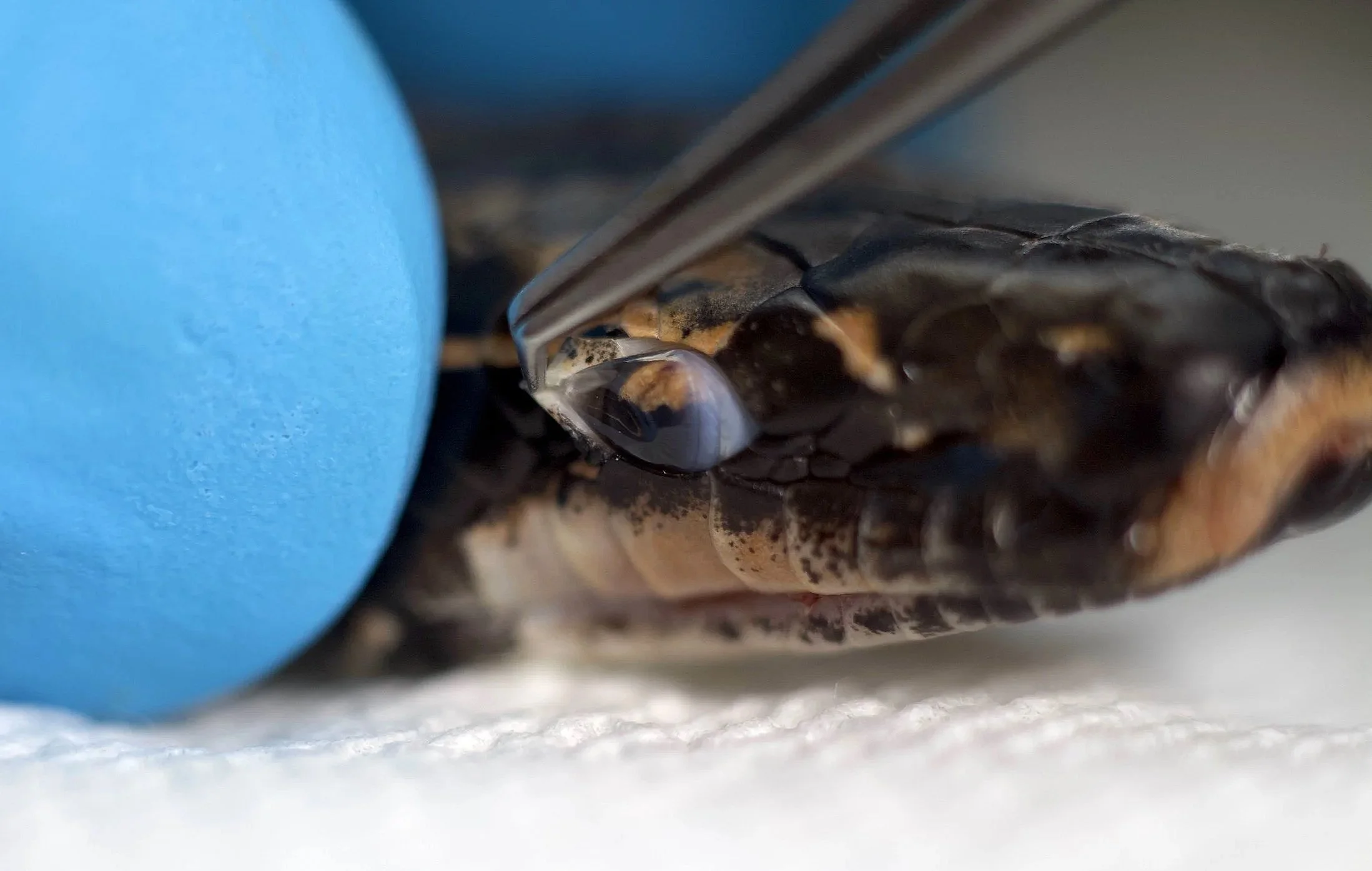
This particular female presented AES on both eyes, with the left eye covered by a large
white scale extending about 70 percent into the spectacle surface. In addition to the large AES, the
postocular scale had an aberrant shape and did not “respect” the orbital border, creating a supra-position in relation to the orbit. (See fig. 7).
The right eye did not have an abnormal white scale over the spectacle, but shared similar
characteristics in relation to the postocular scale extending beyond the proper anatomical limit
between the eye globe and the circumorbital structures. (Fig. 8)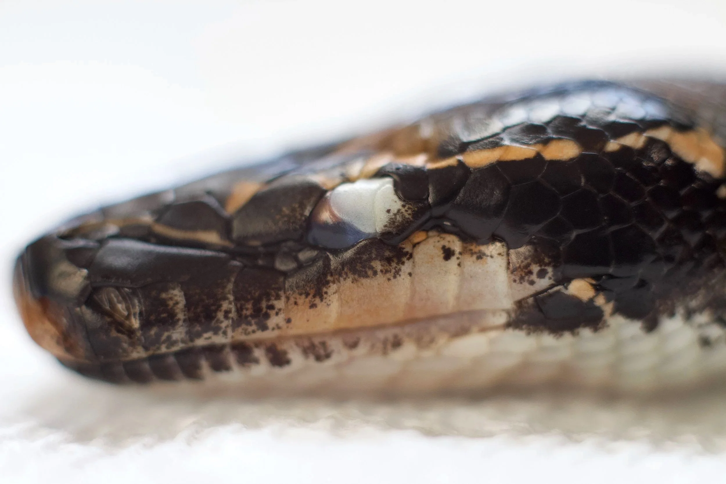
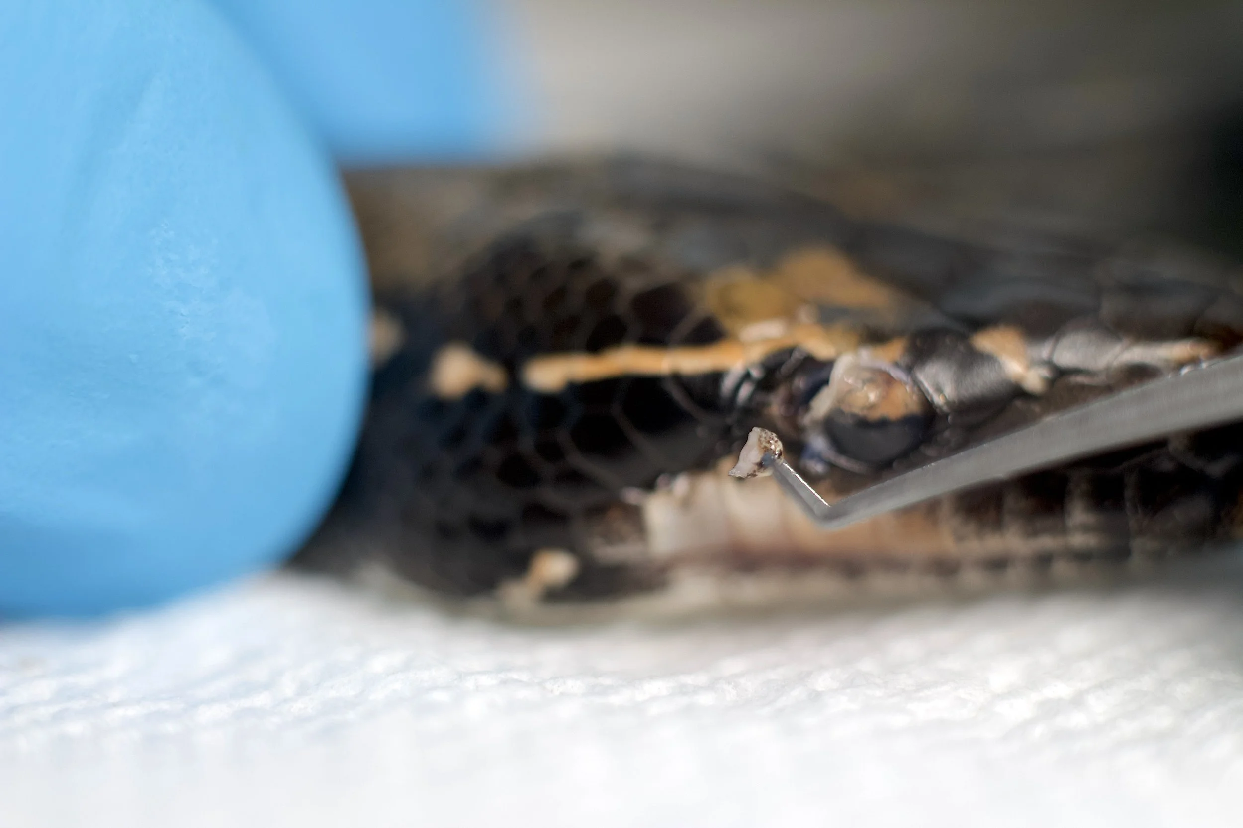
First, I explored the left eye with the largest white scale, using a pair of forceps (colibri) and a
metal hook (Seibel). I followed the edge of the AES, trying to see if the scale was adhering to
the spectacle or not. A closer examination using the metal hook revealed that it was strongly attached to
the spectacle, and it was impossible to penetrate beneath it without an incision. An extension of
clear, hardened keratin extended beyond the white scale into the spectacle, sealing the edges.
Using the forceps, I applied a retracting force to see if the AES would detach easily from the
spectacle by pinching the central portion, as I couldn’t get underneath it (Fig. 9). Instead of detaching, it
moved the entire eyeball following the direction of the forceps. At this point, it was evident that
the AES covering the spectacle was firmly attached to the surface, and surgical removal would be
necessary. (Fig. 10)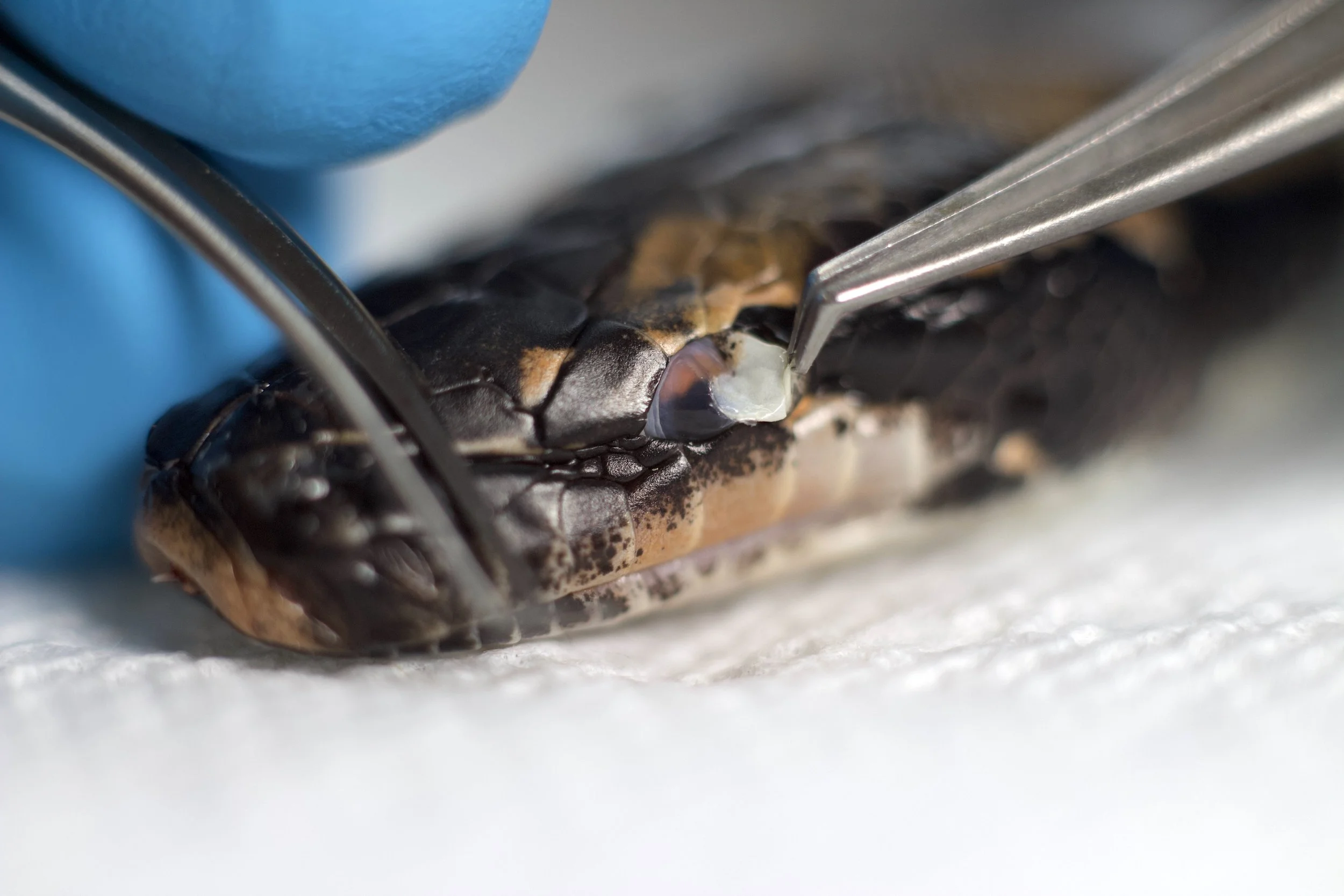
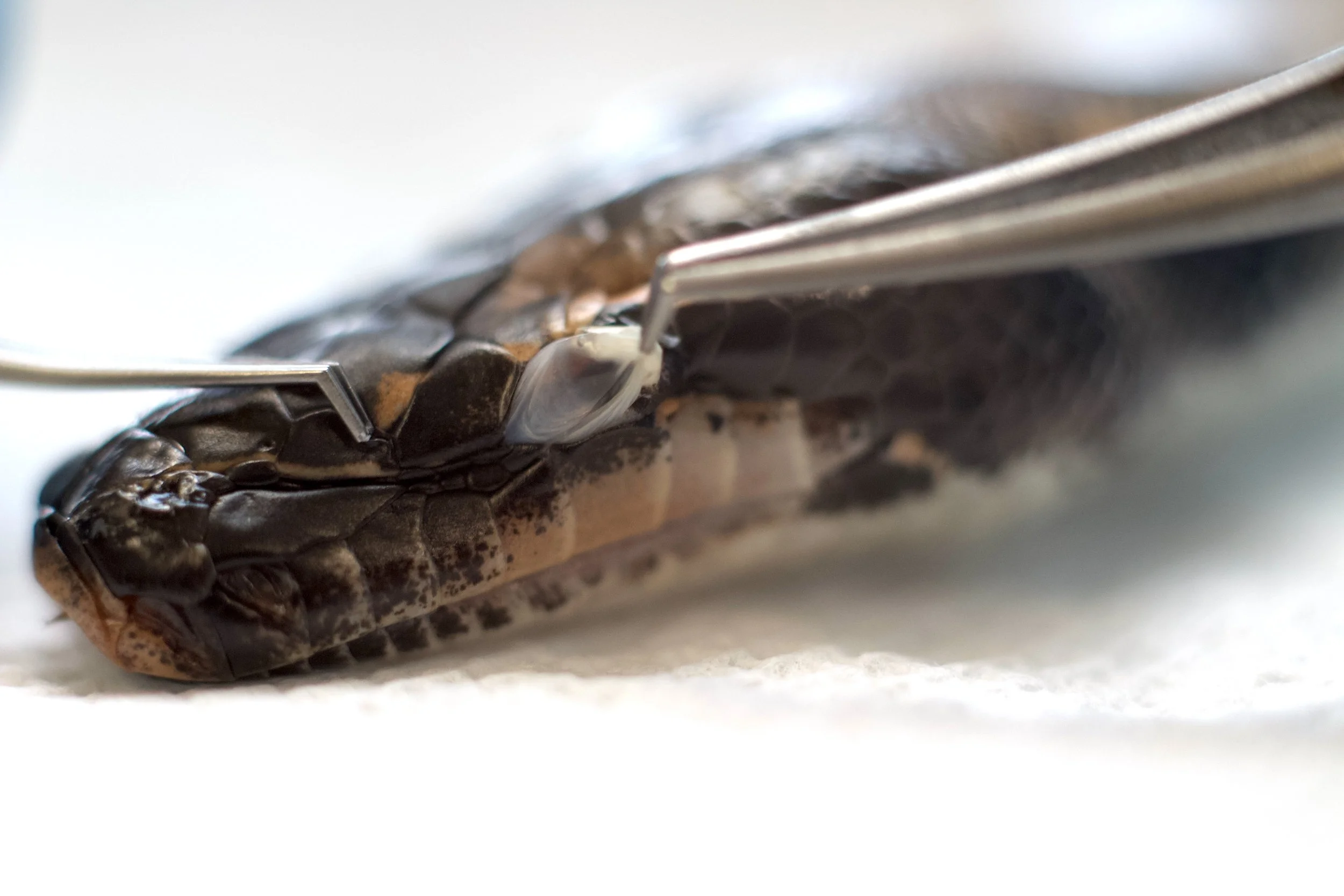
I proceeded to explore the right eye. The abnormal postocular scale had a rounded edge without
the keratin extension, allowing the tip of the hook to go underneath slightly (Fig. 11). The scale was also
firmly adhered to the spectacle, and upon retraction, the entire eyeball moved as well, exposing the
sclera and the connective tissue adhering the spectacle to the inner portion of the postocular scale. This motion revealed that the eyeball inside the ophthalmic chamber was not compromised by the AES, as it could move freely inside the suborbital space, despite the retraction applied to the spectacle, exposing a normal anatomy of the eyeball (Fig. 12). Like with the left eye, a surgical intervention would be necessary to remove the AES. 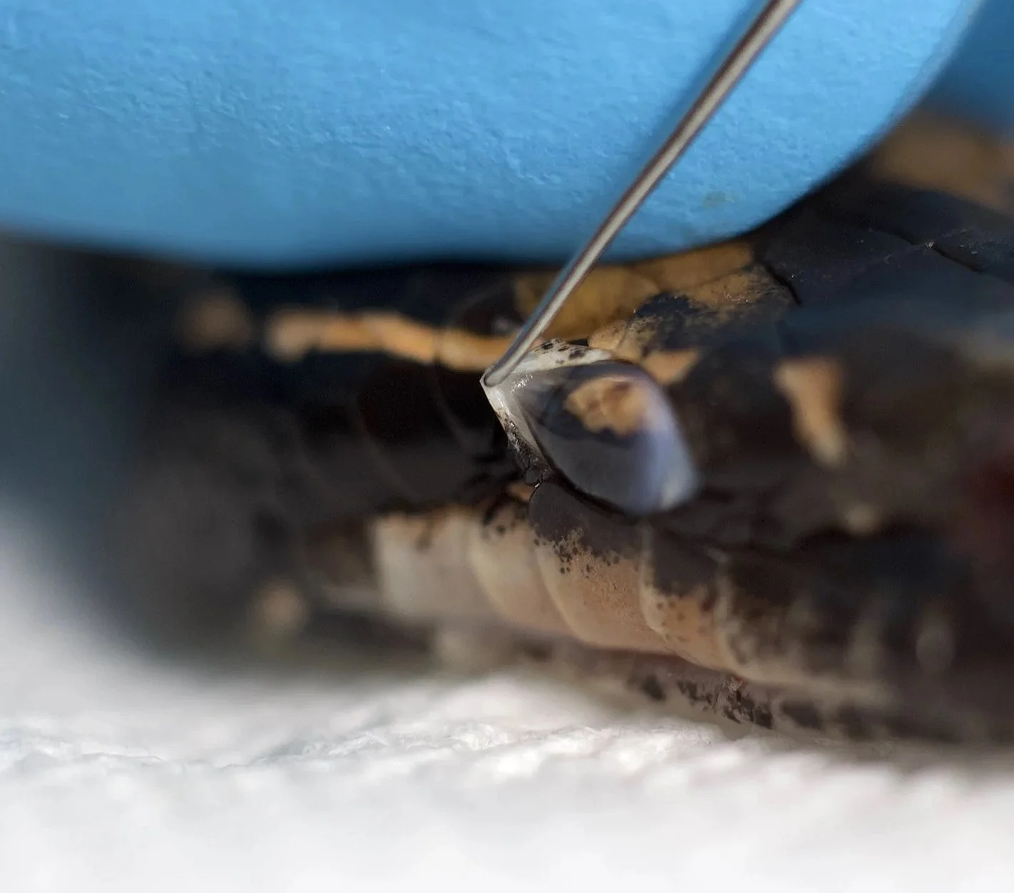
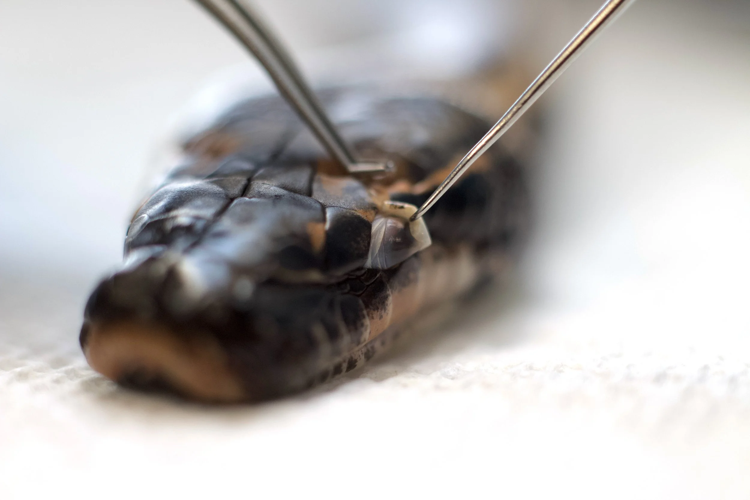
Next, I proceeded to remove the AES. The resection was intended to be delicate and precise, as the goal was to see if removing the abnormal eye scale was possible without damaging the spectacle. Unfortunately, that was not possible. The more I tried, the more I noticed
how intrinsically united both structures were to one another.
The AES was firmly attached to the outer OEG. (Fig. 13).When the AES was finally removed, the portion of the spectacle that was attached to it was also unintentionally removed, exposing the suborbital space and the cornea
beneath it. The portion of the AES closer to the orbit margin contained blood vessels, and
fibrous tissue (Fig. 14), while the AES sitting direclty on the spectacle was a keratin-like structure, similar to a soft fingernail (Fig. 15). The connection between these two structures revealed how hard, if not impossible, it would be to
remove the AES safely without compromising the spectacle integrity and the overall
eyeball health. 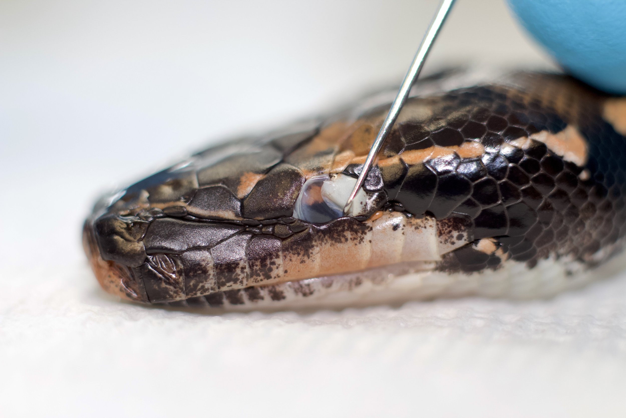
Interestingly, the dissection revealed something fascinating. Despite its appearance, this
“defect” is
entirely superficial and cosmetic. It does not compromise the eyeball itself or other ophthalmic
structures (drainage system, muscles, nerves, and blood vessels). No other specific affections have
been associated with the presence of the AES in WIs, and the Wrought-Irons hatched from non-super WI clutches that have AES are thriving and behaving like normal blood pythons.
Based on these observations, we could say that other than the visual field limitation and cosmetic
implications, the AES does not seem to affect the snakes in any other way. Blood pythons rely on
other adaptations to navigate the world, like their sense of smell and infrared detection abilities,
compensating for the limitations on their visual field. The risks associated with the forced removal
of the AES on snakes affected by it are greater than the benefits, and it is advised to refrain from attempting it, as it could potentially create severe complications for the snake, such as infections or total loss of the eye. Generally
speaking, no special care or treatment is necessary for snakes with AES, as they can thrive and live
healthy, long lives without medical intervention.As our understanding of the AES continues to evolve, collaboration among
breeders will be crucial in uncovering the mechanisms underlying this anomaly. Further studies,
particularly those integrating genetic testing, could provide valuable insights about the AES
and other defects, improving the health and diversity of the Wrought-Iron project. By sharing data
and observations, the community can work together to ensure the well-being of these remarkable
animals and refine best practices for future generations.As always, keep pushing forward! Ernesto Hinojosa